Tendon Adaptations in Throwers: Mechanisms and Applications
Introduction
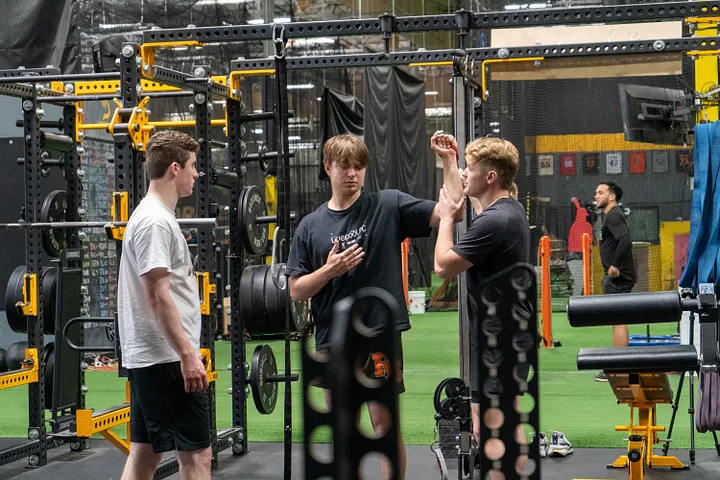
It’s hard to find a more dynamic and physically demanding task in sports than throwing. Whether it be a baseball, javelin, or other implements, high-output throwing tests the boundaries of what the human anatomy is capable of. Throwing is a beautifully orchestrated series of contractions and relaxations across 3D space, analogous to a symphony. The CNS conducts, bones keep the rhythm of percussion, muscles play the brass, and tendons weave the strings. Everyone notices the thunder of the timpani, and it’s impossible to miss the blaring trumpets, but the strings weave those bursts into one seamless chord and let every note ring true.

Tendons are dense, fibrous connective tissues that attach muscles to bones. When muscles contract, tendons pull on bones, producing movement at the joints. Tendons play an important role in throwing, transferring large forces between muscle and bone at high speeds and stabilizing joints throughout the throw. Despite their importance, they are frequently overlooked in holistic throwing performance and injury prevention strategies. The repetitive, high stresses placed on tendons can lead to elbow and shoulder injuries when they are not well-adapted to handle the imposed demands of throwing.
Beyond structural adaptations, neurological factors significantly influence tendon stiffness and compliance. Particularly at the muscle-tendon junction (MTJ) — the critical interface between muscle fibers and tendon collagen — CNS control profoundly shapes tendon behavior. Understanding these neuromechanical interactions can provide new ways to optimize training for performance and injury resilience.
Anatomy and Biomechanics of Throwing
Key Anatomical Structures
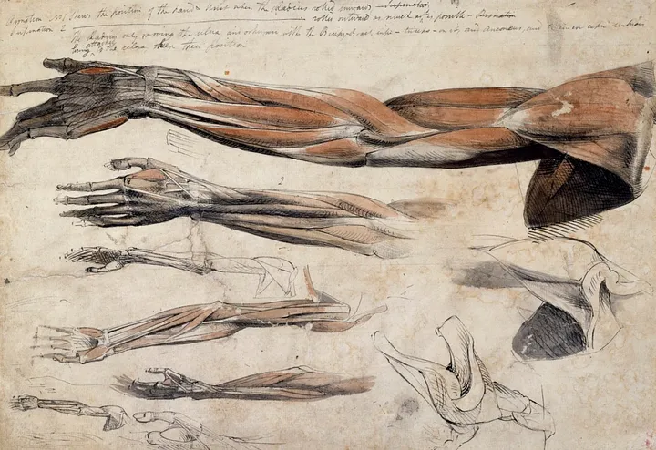
A handful of tendons and ligamentous structures play pivotal roles in supporting the high-speed forces characteristic of the throwing motion. Although this blog focuses on tendon adaptation, the ulnar collateral ligament (UCL) warrants mention because it is a primary stabilizer of the medial elbow. Like tendons, ligaments share many fundamental structural and mechanical properties, enabling them to adapt to progressive loading in broadly similar ways. However, they also exhibit unique structural orientations and functional demands that we won’t go into depth on, emphasizing joint stability rather than force transmission. Despite these differences, the overarching principles of mechanical stress and biological remodeling apply to both tissue types.
Biomechanical Demands
Throwing involves complex interactions among torque, tension, and compression in the throwing arm, resulting in rapid changes in angular velocity and momentum. A primary component of this is valgus torque at the elbow, which places tensile loads on the medial joint structures — particularly the UCL — and compressive forces on the lateral side. Simultaneously, distraction forces pull the humerus away from the scapula, increasing the demands imposed on the rotator cuff and surrounding musculature to maintain proper alignment. The arm also builds up substantial angular momentum during the driveline phase, resulting in extremely high internal rotation torque at the shoulder joint. After ball release, an abrupt deceleration imposes large eccentric loads, requiring the rotator cuff, scapular stabilizers, and other structures to dissipate these forces effectively.

Structural and Mechanical Properties of Tendons
Composition and Hierarchical Organization
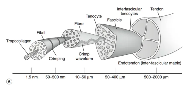
Tendons are complex composite structures primarily composed of collagen, elastin, and proteoglycans. Collagen — particularly Type I — serves as the primary load-bearing element, enabling the tissue to withstand the high forces generated during dynamic movements. Elastin provides the capacity to return to its original shape after stretching, preserving structural integrity through repeated cycles of movement. Proteoglycans help regulate hydration and distribute loads throughout the tendon matrix, enhancing both resilience and flexibility. Beyond these molecular components, tendons exhibit a hierarchical organization: collagen fibrils bundle into fascicles, which are then grouped within larger sheaths and surrounded by the paratenon. This multilayered arrangement equips tendons with exceptional tensile strength and the ability to adapt over time.
Viscoelastic Behavior and Its Importance
A unique quality of tendon mechanics is its viscoelasticity, meaning it exhibits both elastic (spring-like) and viscous (time-dependent) behaviors. In purely elastic materials, stress (σ) scales directly with strain (ϵ) via Hooke’s Law:
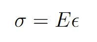
where E (Young’s modulus) indicates stiffness or resistance to deformation. Conversely, purely viscous materials follow Newton’s Law of Viscosity:
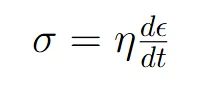
where η represents viscosity. Viscoelastic materials — like tendons — combine these properties, allowing them to store and release energy (elastic response) while also deforming gradually under sustained load (viscous response). As the tendon is stretched, it momentarily resists elongation like a spring, yet over time it can creep or relax, reflecting its viscous component.
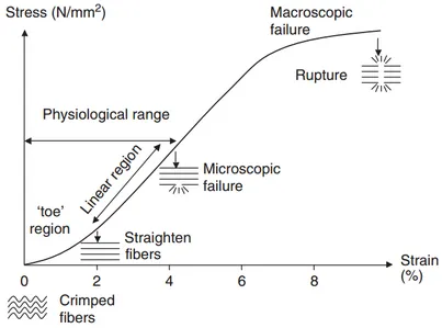
Thickness, Stiffness, and Compliance
Thickness is simply the tendon’s cross-sectional area; add collagen mass and the tendon gets wider, like upgrading a rope from 5 mm to 8 mm. Stiffness is the slope of the stress-strain curve — how many newtons it takes to stretch the tissue by one millimetre. Its inverse is compliance: a highly compliant tendon stretches a lot for a given load, while a very stiff tendon barely budges. Because stiffness is proportional to both the material’s intrinsic elasticity (Young’s modulus) and its thickness, you can thicken a tendon without changing the collagen’s modulus and still end up with a stiffer structure. In practice, thicker tendons distribute stress over more area (protective), whereas stiffer tendons transmit force faster (performance-oriented). Balanced adaptation is therefore about tuning both levers — material quality and size — so the tendon is neither too floppy nor too rigid for the imposed demands.
Understanding Tendon Mechanics
When modeling tendon mechanics, linear viscoelastic approaches (e.g., Maxwell, Kelvin-Voigt, Standard Linear Solid) assume stress and strain remain proportional at moderate loads. However, nonlinear viscoelasticity becomes evident under heavier loads or repetitive high-speed use, where the slope of the tendon’s stress-strain curve flattens with fatigue and structural changes. Several phenomena illustrate these behaviors:
- Creep: Gradual elongation under constant load.
- Stress Relaxation: Decrease in stress when held at a fixed length over time.
- Hysteresis: Loss of energy between loading and unloading cycles.
As living tissues, tendons rely on water diffusion and nutrient transport governed by Fick’s Law, underscoring the biological nature of their adaptation. This continuous influx of nutrients and removal of waste supports collagen turnover and proteoglycan maintenance — both vital for long-term tendon health. Ultimately, their viscoelastic properties make tendons exceptionally well-suited for rapid, high-force activities such as throwing. By briefly deforming and then returning to their original shape, tendons can absorb impact, store elastic energy, and release it efficiently, all while enhancing joint stability. This combination of structural organization and time-dependent mechanics helps tendons remain resilient under the demands of throwing.
The MTJ and Neurological Control
Role and Importance of the MTJ
Crucial to tendon function is the muscle–tendon junction (MTJ), where muscle fibers seamlessly integrate into the collagen fibers of the tendon. This specialized interface transmits muscular forces directly to tendons, undergoing rapid loading cycles during activity. Given its critical role, the MTJ requires precise neurological control to effectively handle rapid, high-amplitude forces.
Central Nervous System Influence
While collagen reorganization and structural remodeling significantly influence tendon adaptations, the central nervous system (CNS) also profoundly affects tendon stiffness through refined neuromuscular control. Specifically, CNS adaptations modify stiffness by regulating muscle activation timing, motor unit recruitment, and the rate and magnitude of muscular force application. These adjustments optimize loading dynamics at the MTJ, effectively reshaping the mechanical responses of tendons.
Motor Unit Recruitment and Synchronization
With repeated high-intensity stimuli — such as plyometric exercises or overcoming isometric holds — the CNS improves its capacity to rapidly recruit and synchronize motor units. This synchronized recruitment intensifies muscular contractions at the MTJ, enhancing the tendon’s elastic response and increasing the efficiency of energy storage and release over time.
Proprioceptive Feedback and Reflexive Control
Muscle spindles and Golgi tendon organs (GTOs) act like two ends of a dial that sets moment-to-moment muscle-tendon unit (MTU) stiffness. Muscle spindles lie in parallel with contractile fibres inside the muscle belly; they fire when those fibres lengthen or lengthen quickly. Their Ia afferents (stretch sensors) monosynaptically excite the same motor-neurons, producing an almost immediate contraction that restores length and adds active stiffness to the system. GTOs, by contrast, sit in series with the fibres inside the tendon at the MTJ. They stay quiet at low load but discharge in proportion to rising tension; their Ib afferents (tension sensors) run through an inhibitory interneuron that tempers the very motor-neurons the spindle just excited, acting as a safety brake when force approaches potentially damaging levels.
Repeated high-speed loading shifts the set-points of both sensors. Spindle gain increases, so pre-activation comes sooner and rate-of-force development climbs, while the GTO threshold drifts upward, allowing greater peak forces before inhibition cuts in. The result is a refined push-pull mechanism: the MTU stiffens precisely when energy must flow from muscle to bone, yet relaxes just in time to protect the structures it’s responsible for.
Neurological Efficiency and Elastic Energy Storage
Repeated exposure to appropriate stimuli refines neuromuscular patterns, optimizing the timing, magnitude, and duration of muscle activation at the MTJ. As the CNS “learns” and consolidates these recruitment patterns, the mechanical efficiency of the MTJ significantly improves, complementing structural collagen adaptations. Neurological adaptations thus collaborate with structural changes to enhance tendon stiffness, ultimately promoting superior elastic energy storage and release during throwing activities.

Cellular and Molecular Mechanisms
Cellular Response to Mechanical Load
At the cellular and molecular levels, tendon adaptation is primarily driven by fibroblasts — often referred to as tenocytes in tendon tissue. Recent research involving fatigue-loaded animal tendons has revealed how mechanical stimuli upregulate several critical genes associated with tissue growth and remodeling. Researchers observed increased expression of collagen types I, III, and V, indicative of anabolic activity and new collagen deposition. Concurrently, MMP-2, MMP-3, and MMP-13 — enzymes responsible for tissue degradation and remodeling — were also elevated, signifying ongoing turnover of damaged tissue. Additionally, TIMP-1 and TIMP-3, proteins involved in inhibiting inflammation, were upregulated, reflecting a balanced interplay between inflammatory and repair pathways in response to mechanical load.
These findings align with previous evidence showing that mechanical load triggers the production of IGF-I, which promotes fibroblast contraction and collagen synthesis. Collectively, the increased activity of collagen, MMP, and TIMP genes highlights the coordinated processes of inflammation, repair, and remodeling that occur following mechanical loading.
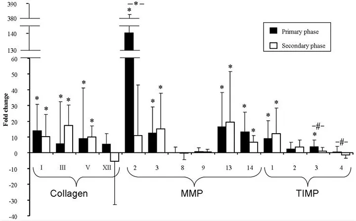
Throughout these stages, maintaining the balance between collagen synthesis and degradation is extremely important. Matrix metalloproteinases (MMPs) remove older or damaged collagen, creating space for new, stronger fibers. However, fibroblast contraction must be tightly regulated — excessive contraction can cause scarring, while insufficient contraction delays healing and compromises tissue quality. Collectively, these cellular activities ensure tendon adaptation occurs in a healthy, progressive manner, ultimately enhancing tendon strength and resilience.
Neuromuscular Influence on Cellular Remodeling
Additionally, neuromuscular signals activated by repetitive loading indirectly influence cellular and molecular pathways. Improved neuromuscular coordination optimizes the timing and distribution of mechanical stresses across the tendon, leading to more uniform cycles of collagen synthesis and degradation. This coordinated signaling ensures even cellular remodeling, reducing localized tissue fatigue and enhancing overall structural integrity.
Structural Adaptations from Stimulus
Positive Structural Changes with Loading
Over time, and with an optimal balance of mechanical loading and recovery, tendons undergo structural adaptations that enhance their ability to manage repetitive, high-speed demands. One of the most observable adaptations is increased tendon thickness, evident in well-conditioned athletes compared to younger or less-trained counterparts. This enlarged cross-sectional area disperses stress across a broader region, facilitating improved load distribution.
Accompanying these dimensional changes are enhancements in mechanical properties, including increased stiffness, greater energy storage capacity, and more efficient force transfer — all of which collectively enhance performance and reduce injury risk. At the microscopic level, collagen fibrils become more densely packed, highly organized, and strongly cross-linked. These adaptations result in a tendon that is simultaneously stiffer and more elastic under heavy or repetitive loads, yet sufficiently compliant to stretch as needed.
Negative Consequences of Tendon Disuse or Immobilization
Conversely, disuse or immobilization can lead to negative adaptations. Prolonged inactivity can induce a net catabolic state within the tendon, characterized by decreased collagen synthesis and accelerated degradation. These changes lead to reduced tendon thickness, diminished stiffness, and potential disorganization of collagen fibrils, ultimately impairing the tissue’s load-bearing capacity. Additionally, prolonged absence of mechanical stimuli may adversely affect vascularization, nutrient transport, and cellular activity, increasing the risk of injury upon the resumption of activity.
Impulse and Loading Dynamics
Understanding Impulse
Impulse refers to the change in momentum, which is the product of force and time elapsed or “duration” (J=Δp=F⋅Δt). As a result, the same impulse can be achieved through different combinations of these two variables. For instance, a large amount of force exerted over a short duration can yield the same overall impulse as a smaller force applied for a longer duration.
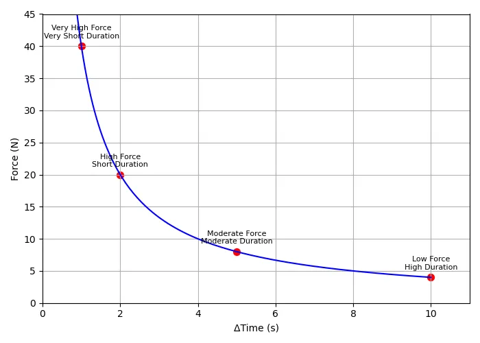
However, the force-time curve — which illustrates how quickly force develops and how long it is sustained — can look drastically different in each scenario.
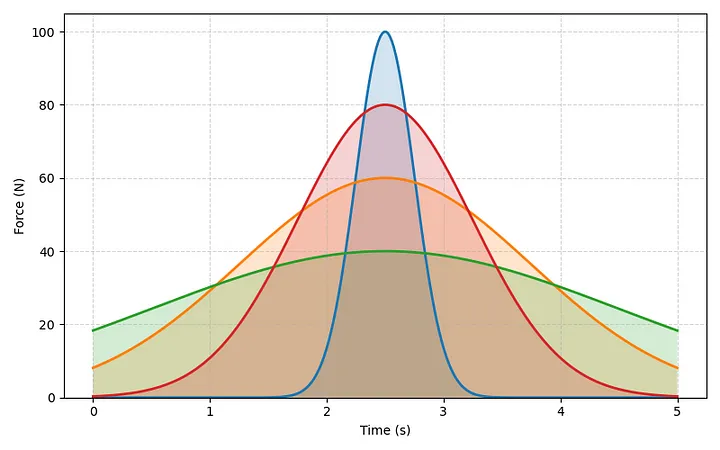
Force-Time Profiles Applied to Tendons
The shape of the force-time curve influences tissue deformation patterns, ultimately resulting in distinct biological responses within the tendon. A rapid spike in force requires the tendon to store and release energy within fractions of a second, stimulating increased stiffness and efficiency at the MTJ. Conversely, a prolonged loading phase emphasizes greater time under tension, driving adaptations such as increased tendon thickness and collagen realignment, but with greater overall deformation. By carefully considering how force changes over time, specific loading patterns can be selected to target desired tendon adaptations effectively.
If you want to learn more about impulse, read Brice Crider’s “Impulse: The King of Performance Metrics.”
Training Stimulus Variables
Impact of Force Magnitude and Loading Duration
Both the magnitude of force and the duration of loading significantly influence tendon adaptations. High peak forces applied within short intervals highlight the tendon’s viscoelastic characteristics, promoting increased stiffness and neuromuscular adaptations crucial for rapid loading and efficient force transmission. In contrast, lower forces applied over longer durations help establish a more robust structural foundation, encouraging tendon thickening and enhancing overall resilience. Together, short-duration loading concentrates high force into a narrow time frame — intensifying the tendon’s demand for elasticity — while prolonged loading phases offer a gradual stimulus that facilitates tissue remodeling and collagen realignment.
Manipulating Frequency and Volume
Adjusting frequency (how often loading sessions occur) and volume (total load per session or training cycle) provides another avenue for guiding tendon adaptation. A higher-frequency, lower-volume approach can enhance tendon stiffness by enabling more repetitions at or near peak intensity, minimizing fatigue and maintaining high output across sessions. Repeated exposure to high peak forces preserves a sharply peaked force-time curve, improving the tendon’s capacity to manage rapid, high-amplitude loads. However, this strategy requires careful workload management to prevent overreaching. Conversely, a lower-frequency, higher-volume approach promotes tendon thickening by accumulating fatigue across more total repetitions, causing temporary reductions in peak output. This prolonged loading stimulates collagen deposition and increased cross-sectional area, though it also extends recovery timelines.
Neurological Benefits
Additionally, specific training stimuli — particularly those emphasizing rapid force application — drive neurological adaptations by refining proprioceptive feedback loops and motor unit recruitment patterns. These neurological improvements reinforce the mechanical properties of tendons, complementing structural changes and further promoting enhanced stiffness.
Neither method is inherently superior; but by fine-tuning these variables — force, time, frequency, and volume — it becomes possible to intentionally elicit specific tendon adaptations through training. If you want to learn more about fatigue management and autoregulation applied to throwing, read Clayton Thompson’s “Understanding and Managing Training Fatigue: From Theory to Practice.”
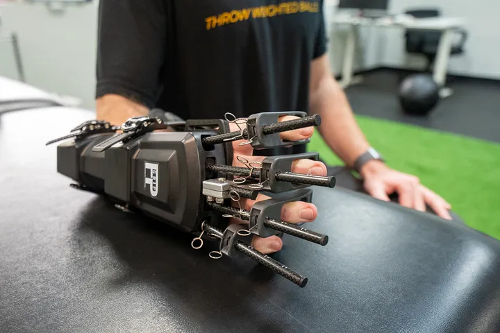
Throwing Implement Selection
Overload Implements: Promoting Structural Robustness
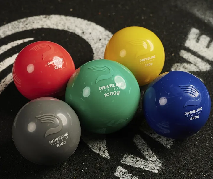
Heavier implements (e.g., overload weighted balls, javelins, footballs) require athletes to generate force over slightly longer durations. Although release velocities are lower compared to lighter implements, the greater mass necessitates substantial force production and thus demands larger impulses to change the implement’s momentum. From a tendon perspective, this prolonged loading promotes structural adaptations, such as increased thickness and enhanced collagen cross-linking, resulting in a robust tendon capable of withstanding repetitive overhead stress without compromising tissue integrity.
Underload Implements: Enhancing Elasticity and Stiffness
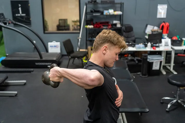
Lighter implements (e.g., underload weighted baseballs, tennis balls) produce higher release velocities for the same impulse, involving higher peak forces over shorter durations. This brief but intense spike on the force-time curve emphasizes the tendon’s elastic properties. Repeated exposure to rapid, high-peak loads promotes increased tendon stiffness and enhances neuromuscular efficiency at the MTJ, improving its capacity to store and release elastic energy. These adaptations can translate directly into greater throwing velocities and improved reactive strength.
Programming Considerations
Strategic Manipulation of Implement Weight
Underload implements capitalize on the tendon’s viscoelastic properties by transmitting force rapidly while inducing comparatively less fatigue than overload implements at equivalent intensities. This reduced fatigue allows athletes to perform high-intensity throwing sessions more frequently. Such an approach is particularly valuable in the late offseason or during in-season training, when maintaining or increasing throwing output is crucial without accumulating unnecessary fatigue. In contrast, overload implements involve prolonged loading durations and, consequently, greater fatigue. Thus, they are better suited to the early offseason, when maximizing absolute output is less critical, and the focus is on enhancing tendon thickness and establishing a robust structural foundation without exposing the tendon to the extreme peak forces associated with lighter implements. This strategy is especially beneficial during return-to-throw or on-ramp programs, as athletes coming off periods without throwing stimulus often have atrophied tendons and reduced MTJ efficiency.
Accomodating Different Training Ages
For less advanced throwers, a mixed-implement approach using both heavy and light implements is generally sufficient. Due to their lower training age and limited adaptations, they benefit from a comprehensive stimulus, promoting simultaneous increases in tendon thickness and stiffness. Varying force magnitude and loading duration within sessions creates diverse loading profiles, preventing overemphasis on any single force-time pattern. In contrast, elite throwers with extensive training history and highly adapted tendons may require a more polarized approach to induce further adaptations in their already advanced qualities. Beginning with an overload-focused strategy and gradually transitioning toward underload training provides dedicated periods at each end of the spectrum, effectively stimulating targeted improvements in tendon properties.
Periodizing Adaptations By Changing Variables
Even without altering implement weight, manipulating volume, intensity, and frequency can effectively drive tendon adaptations. Higher-volume training accumulates stress on the tendon, establishing a foundational level of throwing capacity by gradually enhancing collagen thickness and overall resilience — similar to overload implement training. Conversely, higher-intensity throws generate greater peak forces, prompting the tendon to increase stiffness and improve its rapid force-transfer capability, echoing adaptations typically associated with underload implements.
As athletes progress through a training program, frequency becomes another critical variable — particularly close to competition, when increasing throwing frequency at sustained high intensities helps prepare tendons for repeated sport-specific demands. On shorter timescales, structured models such as high-low programming or undulating periodization provide effective methods for balancing targeted workloads and recovery. By alternating intense throwing sessions with lower-intensity recovery days, the tendon receives the necessary high-stress stimuli alongside essential downtime, facilitating supercompensation once accumulated training fatigue dissipates. This cyclical approach — strategically balancing volume, intensity, and frequency — optimizes tendon health and enhances performance from foundational training phases through competition.
The Role of Submaximal Training
Submaximal effort training differs from maximal effort training in terms of impulse, velocity, and resulting fatigue. Specifically, submaximal throws involve lower velocities and produce less fatigue per throw, allowing athletes to accumulate greater total throwing volume within a session or training cycle. When submaximal effort is combined with overload implements, athletes can achieve higher total tendon stress due to increased cumulative volume, further promoting tendon thickness adaptations. Conversely, applying submaximal training with underload implements enables athletes to train at higher velocities and outputs more frequently, accruing less fatigue and effectively enhancing both tendon thickness and neuromuscular adaptations.
Supplemental Training
Isometric exercises, particularly overcoming isometrics, heavily recruit motor units simultaneously, fostering neurological adaptations that enhance overall MTU response. The maximal voluntary contractions characteristic of these exercises strengthen CNS pathways, refining neuromuscular control and improving tendon efficiency at the MTJ.
Plyometric training also strongly activates proprioceptors, improving muscle spindle and Golgi tendon organ responsiveness. Over repeated cycles, this proprioceptive refinement sharpens reflexive muscle activations, supporting rapid and precise stiffness adjustments crucial for dynamic tendon performance.
Progression and Integration Into Programming
Regardless of the specific exercise, adjustments in duration, velocity, complexity, or effort dictate how tendons will adapt. In isometric training, adaptations may be influenced by longer holds, increased external resistance, or transitioning from yielding to overcoming variations. In plyometric exercises, raising the starting height, reducing ground contact time, or incorporating more open-chain movements can elevate tendon loading and stress.
Ultimately, no exercise for throwers surpasses throwing itself. However, strategically integrating isometric and plyometric methods into a periodized throwing program can help develop thicker, stiffer tendons that are better equipped to withstand the rapid, repetitive forces found in throwing.
Conclusion
By understanding the relationships among force application, tendon structure, and the cellular mechanisms governing adaptation, key tendon qualities — such as thickness, stiffness, and overall load capacity — can be deliberately developed to match the demands of throwing. Adjusting training variables like implement weight, volume, intensity, frequency, and supplementary exercises provides a powerful toolkit for facilitating healthy, progressive tendon remodeling. Furthermore, recognizing the interplay between structural tendon adaptations and neurological improvements — particularly at the muscle–tendon junction — allows athletes and coaches to strategically enhance tendon stiffness and elasticity, ultimately improving performance and injury resilience.
Too many people clean the brass and polish the percussion but neglect the precise tuning that brings the strings into perfect pitch. Treat your tendons as first-chair violinists, and the whole symphony will come to life.
References
[1] Philharmonia. (n.d.) Wilhelm Furtwangler Conducts Philharmonia, Royal Albert Hall, 1947 [Image]. Philharmonia. Retrived from https://philharmonia.co.uk/who-we-are/our-history/
[2] Royal Academy of Arts. (n.d.). A sheet of anatomical drawings of the bones, muscles, and tendons of the arm [Image]. Royal Academy of Arts. Retrieved from https://www.royalacademy.org.uk/art-artists/work-of-art/a-sheet-of-anatomical-drawings-of-the-bones-muscles-and-tendons-of-the-arm
[3] Physio-Pedia. (n.d.). Tendon structure [Image]. Physio-Pedia. Retrieved from https://www.physio-pedia.com/File:/Tendon-structure.png
[4] Physio-Pedia. (n.d.). Tendon stress strain curve [Image]. Physio-Pedia. Retrieved from https://www.physio-pedia.com/File:Tendon-Stress-Strain-Curve.png
[5] Benjamin, M., & Ralphs, J. R. (1998). Fibrocartilage in tendons and ligaments — An adaptation to compressive load. Journal of Anatomy, 193(4), 481–494. https://pubmed.ncbi.nlm.nih.gov/22227881/
[6] Jones, B., Tonniges, J. R., Debski, A., Albert, B., Yeung, D. A., Gadde, N., Mahajan, A., Sharma, N., Calomeni, E. P., Go, M. R., Hans, C. P., & Agarwal, G. (2020). Morphological and biomechanical effects of elastin degradation in the aortic wall. Journal of Biomechanics, 112, 110042. https://www.sciencedirect.com/science/article/abs/pii/S1742706120302191
Comment section